3D / 4D Fetal medicine
Ultrasound and Color Doppler studies
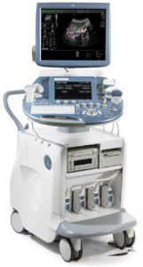
PD boasts of a very high resolution computerized sonographic systems like GE Voluson E8-Radiance and Philips Affinity 70. Apart from routine imagining, these machines add new dimension in volume ultrasound with exceptional anatomical realism and increase depth perception.
Special Studies:
- HD Live Sono-Fetoscopy.
- Early trimester TIFFA (12 to 15 weeks).
- Advance fetal cardiac assessment with high density live 4D.
- High resolution 4D Endovaginal probe (TVS).
- 3D, 4D live TIFFA scan.
- Volume Computer assisted diagnosis of Fetal Heart
- Automated Follicular assessment
- Liver & Breast Elastography
What is 3D ultrasound?
- An ultrasound scan that allows a vision of the fetus in three dimensions.
What is 4D ultrasound?
- A real-time 3D ultrasound and therefore with movement.
- Images and videos obtained by 3D and 4D ultrasound can be recorded for later viewing.
What is the added value of 3D / 4D ultrasound?
- It allows to obtain images of high quality and realism that are usually easier to interpret for parents.
- It has a primarily aesthetic value.
In which cases it is indicated?
- From a medical point of view it is used to improve the study of certain fetal problems.
- In addition, it can be done in a normal pregnancy on parents’ request.
When is it best performed during pregnancy?
- It can be made at any time after 12 weeks of gestation.
- If you only want to do a session, the ideal time is around 28-30 weeks of pregnancy

